Tampilkan postingan dengan label vmPFC. Tampilkan semua postingan

The Brain's Sarcasm Centre? Wow, That's Really Useful

A team of Japanese scientists have found the most sarcastic part of the brain known to date. They also found the metaphor centre of the brain and, well, it's kind of like a pair of glasses.

The paper is Distinction between the literal and intended meanings of sentences and it's brought to you by Uchiyama et al. They took 20 people and used fMRI to record neural activity while the volunteers read 4 kinds of statements:

  • Literally true
  • Nonsensical
  • Sarcastic
  • Metaphorical
The neat thing was that the statements themselves were the same in each case. The preceding context determined how they were to be interpreted. So for example, the statement "It was bone-breaking" was literally true when it formed part of a story about someone in hospital describing an accident; it was metaphorical in the context of someone describing how hard it was to do something difficult; and it was nonsensical if the context was completely unrelated ("He went to the bar and ordered:...").

Here's what they found. Compared to the literally-true and the nonsensical statements, which were a control condition, metaphorical statements activated the head of the caudate nucleus, the thalamus, and an area of the medial PFC they dub the "arMPFC" but which other people might call the pgACC or something even more exotic; names get a bit vague in the frontal lobe.


The caudate nucleus, as I said, looks like a pair of glasses. Except without the nose bit. The area activated by metaphors was the "lenses". Kind of.

Sarcasm however activated the same mPFC region, but not the caudate:

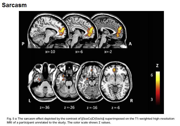
Sarcasm also activated the amygdala.

*

So what? This is a very nice fMRI study. 20 people is a lot, the task was well-designed and the overlap of the mPFC blobs in the sarcasm-vs-control and the metaphor-vs-control tasks was impressive. There's clearly something going on there in both cases, relative to just reading literal statements. Something's going on in the caudate and thalamus with metaphor but not sarcasm, too.

But what can this kind of study tell us about the brain? They've localized something-about-metaphor to the caudate nucleus, but what is it, and what does the caudate actually do to make that thing happen?

The authors offer a suggestion - the caudate is involved in "searching for the meaning" of the metaphorical statement in order to link it to the context, and work out what the metaphor is getting at. This isn't required for sarcasm because there's only one, literal, meaning - it's just reversed, the speaker actually thinks the exact opposite. Whereas with both sarcasm and metaphor you need to attribute intentions (mentalizing or "Theory of Mind").

That's as plausible an account as any but the problem is that we have no way of knowing, at least not from imaging studies, if it's true or not. As I said this is not the fault of this study but rather an inherent challenge for the whole enterprise. The problem is - switch on your caudate, metaphor coming up - a lot like the challenge facing biology in the aftermath of the Human Genome Project.

The HGP mapped the human genome, and like any map it told us where stuff is, in this case where genes are on chromosomes. You can browse it here. But by itself this didn't tell us anything about biology. We still have to work out what most of these genes actually do; and then we have to work out how they interact; and they we have to work out how those interactions interact with other genes and the environment...

Genomics people call this, broadly speaking, "annotating" the genome, although this is not perhaps an ideal term because it's not merely scribbling notes in the margins, it's the key to understanding. Without annotation, the genome's just a big list.

fMRI is building up a kind of human localization map, a blobome if you will, but by itself this doesn't really tell us much; other tools are required.

ResearchBlogging.orgUchiyama HT, Saito DN, Tanabe HC, Harada T, Seki A, Ohno K, Koeda T, & Sadato N (2011). Distinction between the literal and intended meanings of sentences: A functional magnetic resonance imaging study of metaphor and sarcasm. Cortex; a journal devoted to the study of the nervous system and behavior PMID: 21333979

Absolutely Confabulous

Confabulation is a striking symptom of some kinds of brain damage. Patients tell often fantastic stories about things that have happened to them, or that are going on now. It's a classic sign of Korsakoff's syndrome, a disorder caused by vitamin B1 deficiency due to chronic alcoholism.

Korsakoff's was memorably illustrated on House (Season 1 Episode 10, to be exact). Here's a clip; unfortunately, it's overdubbed in Russian, but you can hear the original if you pay attention.

Why does confabulation happen? An influential theory is that confabulation is caused by a failure to filter out irrelevant memories. Suppose I ask you to tell me what happened yesterday. As you reply, yesterday's memories will probably trigger all kinds of associations with other memories, but you'll able to recognize those as irrelevant: that wasn't yesterday, that was last week.

A confabulating patient can't do that, this theory says, so they end up with a huge jumble of memories; the confabulated stories are an attempt to make some sense of this mess. See above for my attempt to confabulate a story linking the three random concepts of a cat, a fire engine and a chair.

Now British neuroscientists Turner, Cipolotti and Shallice argue that this is only part of the truth: Spontaneous confabulation, temporal context confusion and reality monitoring. They discuss three patients, all of whom began to confabulate after suffering ruptured aneurysms of the anterior communicating artery, which destroyed parts of their ventromedial prefrontal cortex.

The patient's stories are tragic, although we can take solace in the fact that they presumably don't know that. The confabulations ranged from the mildly odd:

Patient HS was a 59-year-old man admitted after being found disoriented in the street. [he] had undergone clipping of an ACoA aneurysm 25 years previously. He had been left with a profound confusional state, memory impairment, and confabulation. As a result, HS had been unable to return to work and had spent at least part of the intervening period homeless...

He... continued to produce spontaneous confabulations involving temporal distortions (believing that he had undergone surgery only 18 months previously) and other source memory distortions (confusing memories of interactions with the examiner with interactions with other patients).
To the surreal:
GN was disoriented to place, situation, and time and produced consistent confabulations, for example, believing that the year was 1972 and that he was in a hospital in America after being shot. He regularly produced markedly bizarre confabulations, for example, reporting that he had attended a party the night before and met a woman with a bee’s head. He frequently attempted
to act upon his mistaken beliefs, for example, attempting to leave the hospital to attend meetings.
Anyway, in order to try to discover the mechanism of confabulation, they gave the patients some memory tests. The results were clear: the confabulating patients had no problems remembering stuff, but were unable to tell where they remembered it from.

For example, in one task, the subjects were shown a series of pictures, some of which appeared only once, and some of which were repeated. They had to say which ones were repeats.

The patients did normally the first time they did this task, but when they did the test again, this time with a different subset of pictures repeated, they ran into problems, saying pictures that appeared only once during the session were repeats. They were unable to tell the difference between repeats within the session and repeats from previous sessions. This replicates an earlier study of other confabulators.

But Turner et al found that this lack of awareness for the source of information, wasn't just limited to when things happened. The confabulating patients were also unable to tell the difference between things they'd actually heard, and things they'd only imagined.

Subjects were read a list of 15 words, and also told to silently imagine 15 other words (e.g. "imagine a fruit beginning with A" - apple). They were later asked to remember the words and to say whether they were heard or just imagined. Patients did well on the task except that they wrongly said that they'd actually heard many of the imagined words.

The authors conclude that confabulation is caused by a failure to recognize the source of memories, not just in terms of time, but in terms of whether they were real or fantasy. For a confabulator, all memories are of equal importance. Why this happens as a result of damage to certain parts of the brain remains, however, a mystery.

ResearchBlogging.orgTurner MS, Cipolotti L, & Shallice T (2010). Spontaneous confabulation, temporal context confusion and reality monitoring: A study of three patients with anterior communicating artery aneurysms. Journal of the International Neuropsychological Society : JINS, 1-11 PMID: 20961471

Is Your Brain A Communist?

Capitalists beware. No less a journal than Nature has just published a paper proving conclusively that the human brain is a Communist, and that it's plotting the overthrow of the bourgeois order and its replacement by the revolutionary Dictatorship of the Proletariat even as we speak.

Kind of. The article, Neural evidence for inequality-averse social preferences, doesn't mention the C word, but it does claim to have found evidence that people's brains display more egalitarianism than people themselves admit to.

Tricomi et al took 20 pairs of men. At the start of the study, both men got a $30 payment, but one member of each pair was then randomly chosen to get a $50 bonus. Thus, one guy was "rich", while the other was "poor". Both men then had fMRI scans, during which they were offered various sums of money and saw their partner being offered money too. They rated how "appealing" these money transfers were on a 10 point scale.

What happened? Unsurprisingly both "rich" and "poor" said that they were pleased at the prospect of getting more cash for themselves, the poor somewhat more so, but people also had opinions about payments to the other guy:

the low-pay group disliked falling farther behind the high-pay group (‘disadvantageous inequality aversion’), because they rated positive transfers to the high-pay participants negatively, even though these transfers had no effect on their own earnings. Conversely, the high-pay group seemed to value transfers [to the poor person] that closed the gap between their earnings and those of the low-pay group (‘advantageous inequality aversion’)
What about the brain? When people received money for themselves, activity in the ventromedial prefrontal cortex (vmPFC) and the ventral striatum correlated with the size of their gain.

However, when presented with a payment to the other person, these areas seemed to be rather egalitarian. Activity rose in rich people when their poor colleagues got money. In fact, it was greater in that case than when they got money themselves, which means the "rich" people's neural activity was more egalitarian than their subjective ratings were. Whereas in "poor" people, the vmPFC and the ventral striatum only responded to getting money, not to seeing the rich getting even richer.


The authors conclude that this
indicates that basic reward structures in the brain may reflect even stronger equity considerations than is necessarily expressed or acted on at the behavioural level... Our results provide direct neurobiological evidence in support of the existence of inequality-averse social preferences in the human brain.
Notice that this is essentially a claim about psychology, not neuroscience, even though the authors used neuroimaging in this study. They started out by assuming some neuroscience - in this case, that activity in the vmPFC and the ventral striatum indicates reward i.e. pleasure or liking - and then used this to investigate psychology, in this case, the idea that people value equality per se, as opposed to the alternative idea, that "dislike for unequal outcomes could also be explained by concerns for social image or reciprocity, which do not require a direct aversion towards inequality."

This is known as reverse inference, i.e. inference from data about the brain to theories about the mind. It's very common in neuroimaging papers - we've all done it - but it is problematic. In this case, the problem is that the argument relies on the idea that activity in the vmPFC and ventral striatum is evidence for liking.

But while there's certainly plenty of evidence that these areas are activated by reward, and the authors confirmed that activity here correlated with monetary gain, that doesn't mean that they only respond to reward. They could also respond to other things. For example, there's evidence that the vmPFC is also activated by looking at angry and sad faces.

Or to put it another way: seeing someone you find attractive makes your pupils dilate. If you were to be confronted by a lion, your pupils would dilate. Fortunately, that doesn't mean you find lions attractive - because fear also causes pupil dilation.

So while Tricomi et al argue that people, or brains, like equality, on the basis of these results, I remain to be fully convinced. As Russell Poldrack noted in 2006
caution should be exercised in the use of reverse inference... In my opinion, reverse inference should be viewed as another tool (albeit an imperfect one) with which to advance our understanding of the mind and brain. In particular, reverse inferences can suggest novel hypotheses that can then be tested in subsequent experiments.
ResearchBlogging.orgTricomi E, Rangel A, Camerer CF, & O'Doherty JP (2010). Neural evidence for inequality-averse social preferences. Nature, 463 (7284), 1089-91 PMID: 20182511

Deep Brain Stimulation for Depressed Rats

Deep-brain stimulation (DBS) is probably the most exciting emerging treatment in psychiatry. DBS is the use of high-frequency electrical current to alter the function of specific areas of the brain. Originally developed for Parkinson's disease, over the past five years DBS has been used experimentally in severe clinical depression, OCD, Tourette's syndrome, alcoholism, and more.

Reports of the effects have frequently been remarkable, but there have been few scientifically rigorous studies, and the number of psychiatric patients treated to date is just dozens. So the true usefulness of the technique is unclear. How DBS works is also a mystery. Even the most basic questions - such as whether high-frequency stimulation switches the brain "on" or "off" - are still being debated.

Recent data from rodents sheds some important light on the issue: Antidepressant-Like Effects of Medial Prefrontal Cortex Deep Brain Stimulation in Rats. The authors took rats, and implanted DBS electrodes in the infralimbic cortex. This area is part of the vmPFC. It's believed to be the rat equivalent of the human region BA25, the subgenual cingulate cortex, which is the most common target for DBS in depression. The current settings (100 microA, 130 Hz, 90 microsec) were chosen to be similar to the ones used in humans.

In a standard rat model of depression, the forced-swim test, infralimbic DBS exerted antidepressant-like effects. DBS was equally as effective as imipramine, a potent antidepressant, in terms of reducing "depression-like" behaviours, namely immobility.

This is not all that surprising. Almost everything which treats depression in humans also reduces immobility in this test (along with few things which don't treat it). Much more interesting is what did and did not block the effects of DBS in these rats.

First off, DBS worked even when the rat's infralimbic cortex had been destroyed by the toxin ibotenic acid. This strongly suggests that DBS does not work simply by activating the infralimbic cortex, even though this is where the electrodes were implanted.

Crucially, infralimbic lesions did not have an antidepressant effect per se, which also rules out the theory that DBS works by inactivating this region. (Infralimbic lesions produced by other methods did have a mild antidepressant effect, but it was smaller than the effect of DBS. This may still be important, however.)

What did block the effects of DBS was the depletion of serotonin (5HT). Serotonin is known to its friends as the brain's "happy chemical", although it's a bit more complicated than that. Most antidepressants target serotonin. And rats whose serotonin systems had been lesioned got no benefit from DBS in this study.

So this suggests that DBS might work by affecting serotonin, and indeed, DBS turned out to greatly increase serotonin release, even in a distant part of the brain (the hippocampus). Interestingly this lasted for nearly two hours after the electrodes were switched off.

Depletion of another neurotransmitter, noradrenaline, did not alter the effects of DBS.

Overall, it seems that infralimbic DBS works by increasing serotonin release, but that this is not because it activates or inactivates the infralimbic cortex itself. Rather, nearby structures must be involved. The most likely explanation is that DBS affects nearby white-matter tracts carrying signals between other areas of the brain; the infralimbic cortex might just happen to be "by the roadside". Many researchers believe that this is how DBS works in humans, but this is the first hard evidence for this.

Of course, evidence from rats is never all that hard when it comes to human mental illness. We need to know whether the same thing is true in people. As luck would have it, you can temporarily reduce human serotonin levels with a technique called acute tryptophan depletion This reverses the effects of antidepressants in many people. If this rat data is right, it should also temporarily reverse the benefits of DBS. Someone should do this experiment as soon as possible - I'd like to do it myself, but I'm British, and all the DBS research happens in America. Bah, humbug, old bean.

There's a couple of others things to note here. In other behavioural tests, infralimbic DBS also had antidepressant-like effects: it seemed to reduce anxiety, and it made rats more resistant to the stress of having electrical shocks (although only slightly.) Finally, DBS in another region, the striatum, had no antidepressant effect at all. That's a bit odd because DBS of the striatum does seem to treat depression in humans - but the part of the striatum targeted here, the caudate-putamen, is quite separate to the one targeted in human depression, the nucleus accumbens.

ResearchBlogging.orgHamani, C., Diwan, M., Macedo, C., Brandão, M., Shumake, J., Gonzalez-Lima, F., Raymond, R., Lozano, A., Fletcher, P., & Nobrega, J. (2009). Antidepressant-Like Effects of Medial Prefrontal Cortex Deep Brain Stimulation in Rats Biological Psychiatry DOI: 10.1016/j.biopsych.2009.08.025

 
powered by Blogger