

The search for differences between the brains of men and women has a long and rather confusing history. Any structural differences are small, and their significance is controversial. The one rock-solid finding is that men's brains are slightly bigger on average. Then again, men are slightly bigger on average in general.
A new paper just out from Tomasi and Volkow (of cell-phones-affect-brain fame) offers, on the face of it, extremely strong evidence for a gender difference in the brain, not in structure but in function: Gender Differences in Brain Functional Connectivity Density.
Here's the headline pic: They used resting-state "functional connectivity" (though see here for why this term may be misleading) fMRI in men and women. This essentially means that they put people in the MRI scanner, told them to just lie there and relax, and measured the degree to which activity in different parts of the brain was correlated to activity in every other part. They had a whopping 561 brains in total, though they didn't scan everyone themselves: they downloaded the data from here.
As you can see the results were highly consistent around the world. In both men and women, the main "connectivity hub" was an area called the ventral precuneus. This is interesting in itself although not a new finding as the precuneus has long been known to be involved in resting-state networks. However, the degree of connectivity was higher in women than in men 14% higher, in fact.
The method they used, which they've dubbed "Local Functional Connectivity Density Mapping", is apparantly a fast way of calculating the degree to which each part of the brain is functionally related to each other part.
You could do this by taking every single voxel and correlating it with every other voxel, for every single person, but this would take forever unless you had a supercomputer. LFCDM is, they say, a short-cut. I'm not really qualified to judge whether it's a valid one, but it looks solid.
Also, men's brains were on average bigger, but interestingly they show that women had, relative to brain size, more grey matter than men. Here's the data (I'm not sure about the color scheme...) So what does the functional connectivity finding mean? It could mean anything, or nothing. You could interpret the highly interconnected female brain as an explanation for why women are more holistic, better at multi-tasking, and more in touch with their emotions than men with their fragmented faculties. Or whatever.
Or you could say, that that's sexist rubbish, and all this means is that men and women on average are thinking about different things when they lie in MRI scanners. We already know that resting-state functional connectivity centred on the precuneus is suppressed whenever your attention is directed towards an external "task".
That's not a fault of this research, which is excellent as far as it goes and certainly raises lots of interesting questions about functional connectivity. But we don't know what it means quite yet.
Women Are Better Connected... Neurally
 23.50
23.50
 wsn
wsn
 Posted in
fMRI,
neurofetish,
papers
Posted in
fMRI,
neurofetish,
papers
The Brain's Sarcasm Centre? Wow, That's Really Useful
12.25
wsn
A team of Japanese scientists have found the most sarcastic part of the brain known to date. They also found the metaphor centre of the brain and, well, it's kind of like a pair of glasses. The paper is Distinction between the literal and intended meanings of sentences and it's brought to you by Uchiyama et al. They took 20 people and used fMRI to record neural activity while the volunteers read 4 kinds of statements:
- Literally true
- Nonsensical
- Sarcastic
- Metaphorical
Here's what they found. Compared to the literally-true and the nonsensical statements, which were a control condition, metaphorical statements activated the head of the caudate nucleus, the thalamus, and an area of the medial PFC they dub the "arMPFC" but which other people might call the pgACC or something even more exotic; names get a bit vague in the frontal lobe.

The caudate nucleus, as I said, looks like a pair of glasses. Except without the nose bit. The area activated by metaphors was the "lenses". Kind of.
Sarcasm however activated the same mPFC region, but not the caudate:
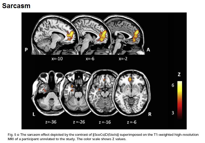 Sarcasm also activated the amygdala.
But what can this kind of study tell us about the brain? They've localized something-about-metaphor to the caudate nucleus, but what is it, and what does the caudate actually do to make that thing happen?
The authors offer a suggestion - the caudate is involved in "searching for the meaning" of the metaphorical statement in order to link it to the context, and work out what the metaphor is getting at. This isn't required for sarcasm because there's only one, literal, meaning - it's just reversed, the speaker actually thinks the exact opposite. Whereas with both sarcasm and metaphor you need to attribute intentions (mentalizing or "Theory of Mind").
That's as plausible an account as any but the problem is that we have no way of knowing, at least not from imaging studies, if it's true or not. As I said this is not the fault of this study but rather an inherent challenge for the whole enterprise. The problem is - switch on your caudate, metaphor coming up - a lot like the challenge facing biology in the aftermath of the Human Genome Project.
The HGP mapped the human genome, and like any map it told us where stuff is, in this case where genes are on chromosomes. You can browse it here. But by itself this didn't tell us anything about biology. We still have to work out what most of these genes actually do; and then we have to work out how they interact; and they we have to work out how those interactions interact with other genes and the environment...
Genomics people call this, broadly speaking, "annotating" the genome, although this is not perhaps an ideal term because it's not merely scribbling notes in the margins, it's the key to understanding. Without annotation, the genome's just a big list.
fMRI is building up a kind of human localization map, a blobome if you will, but by itself this doesn't really tell us much; other tools are required.
Uchiyama HT, Saito DN, Tanabe HC, Harada T, Seki A, Ohno K, Koeda T, & Sadato N (2011). Distinction between the literal and intended meanings of sentences: A functional magnetic resonance imaging study of metaphor and sarcasm. Cortex; a journal devoted to the study of the nervous system and behavior PMID: 21333979Premature Brain Diagnosis in Japan?
15.20
wsn
Nature has a disturbing article from their Asian correspondent David Cyranoski: Thought experiment. It's open access. In brief: a number of top Japanese psychiatrists have started offering a neuroimaging method called NIRS to their patients as a diagnostic tool. They claim that NIRS shows the neural signatures of different mental illnesses.
The technology was approved by the Japanese authorities in April 2009, and since then it's been used on at least 300 patients, who pay $160 for the privilege. However, it's not clear that it works.
To put it mildly.
It's a lot cheaper and easier than MRI. However, the images it provides are a lot less detailed, and it can only image the surface of the brain. NIRS has a small but growing number of users in neuroscience research; it's especially popular in Japan, for some reason, but it's also found plenty of users elsewhere.
The clinical use of NIRS in psychiatry was pioneered by one Dr Masato Fukuda, and he's been responsible for most of the trials. So what are these trials?
As far as I can see (correct me if I'm wrong), these are all the trials comparing patients and controls that he's been an author on:
- Matsuo et al (2000) n=9/10 elderly depressed/controls
- Suto et al (2004) n=10/13/16 depressed/schizophrenia/controls
- Kameyama et al (2006) n=17/11/17 bipolar/depression/controls
- Nishimura et al (2007) n=5/33 panic disorder/controls
- Takizawa et al (2008) n=55/70 schizophrenia/controls
- Uehara et al (2007) n=11/11 eating disorder/controls
- Suda et al (2010) n=27/27 eating disorder/controls
So we have 342 people in all. Actually, a bit less, because some of them were included in more than one study. That's still quite a lot - but there were only 5 panic patients, 30 depressed (including 9 elderly, who may be different), 38 eating disordered and just 17 bipolar in the mix.
And the bipolar people were currently feeling fine, or just a little bit down, at the time of the NIRS. There are quite a lot of other trials from other Japanese groups, but sticking with bipolar disorder as an example, no trials that I could find examined people who were currently ill. The only other two trials, both very small, were in recovered people (1,2).
Given that the whole point of diagnosis is to find out what any given patient has, when they're ill, this matters to every patient. Anyone could be psychotic, or depressed, or eating disordered, or any combination thereof.
Worse yet, in many of these studies the patients were taking medications. In the 2006 depression/bipolar paper, for example, all of the bipolars were on heavy-duty mood stabilizers, mostly lithium; plus a few antipsychotics, and lots of antidepressants. The depressed people were on antidepressants.
There's a deeper problem. Fukuda says that NIRS corresponds with the clinical diagnosis in 80% of cases. Let's assume that's true. Well, if the NIRS agrees with the clinical diagnosis, it doesn't tell us anything we didn't already know. If the NIRS disagrees, who do you trust?
I think you'd have to trust the clinician, because the clinician is the "gold standard" against which the NIRS is compared. Psychiatric diseases are defined clinically. If you had to choose between 80% gold and pure gold, it's not a hard choice.
Now NIRS could, in theory, be better than clinical diagnosis: it could provide more accurate prognosis, and more useful treatment recommendations. That would be cool. But as far as I can see there's absolutely no published evidence on that.
To find out you'd have to compare patients diagnosed with NIRS to patients diagnosed normally - or better, to those randomized to get fake placebo NIRS, like the authors of this trial from last year should have done. To my knowledge, there have been no such tests at all.
 So what? NIRS is harmless, quick, and $160 is not a lot. Patients like it: “They want some kind of hard evidence,” [Fukuda says], especially when they have to explain absences from work. If it helps people to come to terms with their illness - no mean feat in many cases - what's the problem?
My worry is that it could mean misdiagnosing patients, and therefore mis-treating them. Here's the most disturbing bit of the article:
...when Fukuda calculates his success rates, NIRS results that match the clinical diagnosis are considered a success. If the results don’t match, Fukuda says he will ask the patient and patient’s family “repeatedly” whether they might have missed something — for example, whether a depressed patient whose NIRS examination suggests schizophrenia might have forgotten to mention that he was experiencing hallucinations.Quite apart from the implication that the 80% success rate might be inflated, this suggests that some dubious clinical decisions might be going on. The first-line treatments for schizophrenia are quite different, and rather less pleasant, than those for depression. A lot of perfectly healthy people report "hallucinations" if you probe hard enough. "Seek, and ye shall find". So be careful what you seek for.
While NIRS is a Japanese speciality, other brain-based diagnostic or "treatment personalization" tools are being tested elsewhere. In the USA, EEG has been proposed by a number of groups. I've been rather critical of these methods, but at least they've done some trials to establish whether this actually improves patient outcomes.
In my view, all of these "diagnostic" or "predictive" tools should be subject to exactly the same tests as treatments are: double blind, randomized, sham-controlled trials.
Cyranoski, D. (2011). Neuroscience: Thought experiment Nature, 469 (7329), 148-149 DOI: 10.1038/469148a Posted in
bad neuroscience,
mental health,
neurofetish,
papers
Left Wing vs. Right Wing Brains
08.50
wsn
So apparently: Left wing or right wing? It's written in the brain
People with liberal views tended to have increased grey matter in the anterior cingulate cortex, a region of the brain linked to decision-making, in particular when conflicting information is being presented...
Conservatives, meanwhile, had increased grey matter in the amygdala, an area of the brain associated with processing emotion.
Politics blog Heresy Corner discusses it...
Subjects who professed liberal or left-wing opinions tended to have a larger anterior cingulate cortex, an area of the brain which, we were told, helps process complex and conflicting information. (Perhaps they need this extra grey matter to be able to cope with the internal contradictions of left-wing philosophy.)
In truth, without seeing the full scientific paper, we can't know whether the differences they found were really statistically solid, or whether they were voodoo or fishy. The authors, Geraint Rees and Ryota Kanai, have both published a lot of excellent neuroscience in the past, but that's no guarantee.
In fact, however, I suspect that the brain is just the wrong place to look if you're interested in politics, because most political views don't originate in the individual brain, they originate in the wider culture and are absorbed and regurgitated without much thought. This is a real shame, because all of us, left or right, have a brain, and it's really quite nifty:
 But when it comes to politics we generally don't use it. The brain is a powerful organ designed to help you deal with reality in all its complexity. For a lot of people, politics doesn't take place there, it happens in fairytale kingdoms populated by evil monsters, foolish jesters, and brave knights.
 Given that the characters in this story are mindless stereotypes, there's no need for empathy. Because the plot comes fully-formed from TV or a newspaper, there's no need for original ideas. Because everything is either obviously right or obviously wrong, there's not much reasoning required. And so on. Which is why this happens amongst other things.
I don't think individual personality is very important in determining which political narratives and values you adopt: your family background, job, and position in society is much more important.
Where individual differences matter, I think, is in deciding how "conservative" or "radical" you are within whatever party you find yourself. Not in the sense of left or right, but in terms of how keen you are on grand ideas and big changes, as opposed to cautious, boring pragmatism.
In this sense, there are conservative liberals (i.e. Obama) and radical conservatives (i.e. Palin), and that's the kind of thing I'd be looking for if I were trying to find political differences in the brain.
Links: If right wingers have bigger amygdalae, does that mean patient SM, the woman with no amygdalae at all, must be a communist? Then again, Neuroskeptic readers may remember that the brain itself is a communist...
Posted in
blogging,
controversiology,
media,
neurofetish,
politics
Delusions of Gender
13.50
wsn
Note: This book quotes me approvingly, so this is not quite a disinterested review.
Cordelia Fine's Delusions of Gender is an engaging, entertaining and powerfully argued reply to the many authors - who range from the scientifically respectable to the less so - who've recently claimed to have shown biological sex differences in brain, mind and behaviour. Fine makes a strong case that the sex differences we see, in everything from behaviour to school achievements in mathematics, could be caused by the society in which we live, rather than by biology. Modern culture, she says, while obviously less sexist than in the past, still contains deeply entrenched assumptions about how boys and girls ought to behave, what they ought to do and what they're good at, and these - consciously or unconsciously - shape the way we are.
Some of the Fine's targets are obviously bonkers, like Vicky Tuck, but for me, the most interesting chapters were those dealing in detail with experiments which have been held up as the strongest examples of sex differences, such as the Cambridge study claiming that newborn boys and girls differ in how much they prefer looking at faces as opposed to mechanical mobiles.
But Delusions is not, in Steven Pinker's phrase, saying we ought to return to "Blank Slatism", and it doesn't try to convince you that every single sex difference definately is purely cultural. It's more modest, and hence, much more believable: simply a reminder that the debate is still an open one.
Fine makes a convincing case (well, it convinced me) that the various scientific findings, mostly from the past 10 years, that seem to prove biological differences, are not, on the whole, very strong, and that even if we do accept their validity, they don't rule out a role for culture as well.
This latter point is, I think, especially important. Take, for example, the fact that in every country on record, men roughly between the ages of 16-30 are responsible for the vast majority of violent crimes. This surely reflects biology somehow; whether it's the fact that young men are physically the strongest people, or whether it's more psychological, is by the by.
But this doesn't mean that young men are always violent. In some countries, like Japan, violent crime is extremely rare; in other countries, it's tens of times more common; and during wars or other periods of disorder, it becomes the norm. Young men are always, relatively speaking, the most violent but the absolute rate of violence varies hugely, and that has nothing to do with gender. It's not that violent places have more men than peaceful ones.
Gender, in other words, doesn't explain violence in any useful way - even though there surely are gender differences. The same goes for everything else: men and women may well have, for biological reasons, certain tendencies or advantages, but that doesn't automatically explain (and it doesn't justify) all of the sex differences we see today; it's only ever a partial explanation, with culture being the other part.
Posted in
autism,
bad neuroscience,
books,
neurofetish
Genes To Brains To Minds To... Murder?
00.22
wsn
A group of Italian psychiatrists claim to explain How Neuroscience and Behavioral Genetics Improve Psychiatric Assessment: Report on a Violent Murder Case. The paper presents the horrific case of a 24 year old woman from Switzerland who smothered her newborn son to death immediately after giving birth in her boyfriend's apartment. After her arrest, she claimed to have no memory of the event. She had a history of multiple drug abuse, including heroin, from the age of 13.
But that's not all. In the paper, the authors bring neuroscience and genetics into the case in an attempt to provide
a more “objective description” of the defendant’s mental disease by providing evidence that the disease has “hard” biological bases. This is particularly important given that psychiatric symptoms may be easily faked as they are mostly based on the defendant’s verbal report.So they scanned her brain, and did DNA tests for 5 genes which have been previously linked to mental illness, impulsivity, or violent behaviour. What happened? Apparently her brain has "reduced gray matter volume in the left prefrontal cortex" - but that was compared to just 6 healthy control women. You really can't do this kind of analysis on a single subject, anyway.
As for her genes, well, she had genes. On the famous and much-debated 5HTTLPR polymorphism, for example, her genotype was long/short; while it's true that short is generally considered the "bad" genotype, something like 40% of white people, and an even higher proportion of East Asians, carry it. The situation was similar for the other four genes (STin2 (SCL6A4), rs4680 (COMT), MAOA-uVNTR, DRD4-2/11, for gene geeks).
I've previously posted about cases in which a well-defined disorder of the brain led to criminal behaviour. There was the man who became obsessed with child pornography following surgical removal of a tumour in his right temporal lobe. There are the people who show "sociopathic" behaviour following fronto-temporal degeneration.
However this woman's brain was basically "normal" at least as far as a basic MRI scan could determine. All the pieces were there. Her genotypes was also normal in that lots of normal people carry the same genes; it's not (as far as we know) that she has a rare genetic mutation like Brunner syndrome in which an important gene is entirely missing. So I don't think neurobiology has much to add to this sad story.
There seems to be a basic difference between the way in which we think about "biological" as opposed to "environmental" causes of behaviour. This is related, I think, to the Seductive Allure of Neuroscience Explanations and our fascination with brain scans that "prove that something is in the brain". But when you start to think about it, it becomes less and less clear that this distinction works.
A person's family, social and economic background is the strongest known predictor of criminality. Guys from stable, affluent families rarely mug people; some men from poor, single-parent backgrounds do. But muggers don't choose to be born into that life any more than the child-porn addict chose to have brain cancer.
Indeed, the mugger's situation is a more direct cause of his behaviour than a brain tumour. It's not hard to see how a mugger becomes, specifically, a mugger: because they've grown up with role-models who do that; because their friends do it or at least condone it; because it's the easiest way for them to make money.
 But it's less obvious how brain damage by itself could cause someone to seek child porn. There's no child porn nucleus in the brain. Presumably, what it does is to remove the person's capacity for self-control, so they can't stop themselves from doing it.
This fits with the fact that people who show criminal behaviour after brain lesions often start to eat and have (non-criminal) sex uncontrollably as well. But that raises the question of why they want to do it in the first place. Were they, in some sense, a pedophile all along? If so, can we blame them for that?
Rigoni D, Pellegrini S, Mariotti V, Cozza A, Mechelli A, Ferrara SD, Pietrini P, & Sartori G (2010). How neuroscience and behavioral genetics improve psychiatric assessment: report on a violent murder case. Frontiers in behavioral neuroscience, 4 PMID: 21031162 Posted in
ethics,
genes,
law,
neurofetish,
papers,
philosophy
Brain Scans Prove That The Brain Does Stuff
14.01
wsn
The US scientists behind the study suggest it provides solid evidence that the problem can have a physical origin. According to the BBC (and many others)...
Libido problems 'brain not mind'
The research in question (which hasn't been published yet) has been covered very well over at The Neurocritic. Basically the authors took some women with a diagnosis of "Hypoactive Sexual Desire Disorder" (HSDD), and some normal women, put them in an fMRI scanner and showed them porn. Different areas of the brain lit up.
Scans appear to show differences in brain functioning in women with persistently low sex drives, claim researchers.
So what? For starters we have no idea if these differences are real or not because the study only had a tiny 7 normal women, although strangely, it included a full 19 women with HSDD. Maybe they had difficulty finding women with healthy appetites in Detroit?
Either way, a study is only as big as its smallest group so this was tiny. We're also not told anything about the stats they used so for all we know they could have used the kind that give you "results" if you use them on a dead fish.
But let's grant that the results are valid. This doesn't tell us anything we didn't already know. We know the women differ in their sexual responses - because that's the whole point of the study. And we know that this must be something to do with their brain, because the brain is where sexual responses, and every other mental event, happen.
So we already know that HSDD "has a physical origin", but only in the sense that everything does; being a Democrat or a Republican has a physical origin; being Christian or Muslim has a physical origin; speaking French as opposed to English has a physical origin; etc. etc. None of which is interesting or surprising in the slightest.
The point is that the fact that something is physical doesn't stop it being also psychological. Because psychology happens in the brain. Suppose you see a massive bear roaring and charging towards you, and as a result, you feel scared. The fear has a physical basis, and plenty of physical correlates like raised blood pressure, adrenaline release, etc.
But if someone asks "Why are you scared?", you would answer "Because there's a bear about to eat us", and you'd be right. Someone who came along and said, no, your anxiety is purely physical - I can measure all these physiological differences between you and a normal person - would be an idiot (and eaten).
Now sometimes anxiety is "purely physical" i.e. if you have a seizure which affects certain parts of the temporal lobe, you may experience panic and anxiety as a direct result of the abnormal brain activity. In that case the fear has a physiological cause, as well as a physiological basis.
Maybe "HSDD" has a physiological cause. I'm sure it sometimes does; it would be very weird if it didn't in some cases because physiology can cause all kinds of problems. But fMRI scans don't tell us anything about that.
Link: I've written about HSDD before in the context of flibanserin, a drug which was supposed to treat it (but didn't). Also, as always, British humour website The Daily Mash hit this one on the head...
Posted in
bad neuroscience,
flibanserin,
fMRI,
media,
mental health,
neurofetish,
woo
You're (Brain Is) So Immature
06.30
wsn
How mature are you? Have you ever wanted to find out, with a 5 minute brain scan? Of course you have. And now you can, thanks to a new Science paper, Prediction of Individual Brain Maturity Using fMRI. This is another clever application of the support vector machine (SVM) method, which I've written about previously, most recently regarding "the brain scan to diagnose autism". An SVM is a machine learning algorithm: give it a bunch of data, and it'll find patterns in it.
In this case, the input data was brain scans from children, teenagers and adults, and the corresponding ages of each brain. The pattern the SVM was asked to find was the relationship between age and some complex set of parameters about the brain.
The scan was resting state functional connectivity fMRI. This measures the degree to which different areas of the brain tend to activate or deactivate together while you're just lying there (hence "resting"). A high connectivity between two regions means that they're probably "talking to each other", although not necessarily directly.
It worked fairly well: Out of 238 people aged 7 to 30, the SVM was able to "predict" age pretty nicely on the basis of the resting state scan. This graph shows chronological age against predicted brain age (or "fcMI" as they call it). The correlation is strong: r2=0.55.
The authors then tested it on two other large datasets: one was resting state, but conducted on a less powerful scanner (1.5T vs 3.0T) (n=195), and the other was not designed as a resting state scan at all, but did happen to include some resting state-like data (n=186). Despite the fact that these data were, therefore, very different to the original dataset, the SVM was able to predict age with r2 over 0.5 as well.
What use would this be? Well, good question. It would be all too easy to, say, find a scan of your colleague's brain, run it through the Mature-O-Meter, and announce with glee that they have a neurological age of 12, which explains a lot. For example.
However, while this would be funny, it wouldn't necessarily tell you anything about them. We already know everyone's neurological age. It's... their age. Your brain is an old as you are. These data raise the interesting possibility that people with a higher Maturity Index, for their age, are actually more "mature" people, whatever that means. But that might not be true at all. We'll have to wait and see.
How does this help us to understand the brain? An SVM is an incredibly powerful mathematical tool for detecting non-linear correlations in complex data. But just running an SVM on some data doesn't mean we've learned anything: only the SVM has. It's a machine learning algorithm, that's what it does. There's a risk that we'll get "science without understanding" as I've written a while back.
In fact the authors did make a start on this and the results were pretty neat. They found that as the brain matures, long-range functional connections within the brain become stronger, but short-range interactions between neighbours get weaker and this local disconnection with age is the most reliable change.
 You can see this on the pic above: long connections get stronger (orange) while short ones get weaker (green), in general. This is true all across the brain.
It's like how when you're a kid, you play with the kids next door, but when you grow up you spend all your time on the internet talking to people thousands of miles away, and never speak to your neighbours. Kind of.
Link: Also blogged about here.
Dosenbach NU, Nardos B, Cohen AL, Fair DA, Power JD, Church JA, Nelson SM, Wig GS, Vogel AC, Lessov-Schlaggar CN, Barnes KA, Dubis JW, Feczko E, Coalson RS, Pruett JR Jr, Barch DM, Petersen SE, & Schlaggar BL (2010). Prediction of individual brain maturity using fMRI. Science (New York, N.Y.), 329 (5997), 1358-61 PMID: 20829489 Posted in
fMRI,
methods,
neurofetish,
papers
Drugs for Starcraft Addiction
15.05
wsn
Are you addicted to Starcraft? Do you want to get off Battle.net and on a psychoactive drug? Well, South Korean psychiatrists Han et al report that Bupropion sustained release treatment decreases craving for video games and cue-induced brain activity in patients with Internet video game addiction.
They took 11 people with "Internet Game Addiction" - the game being Starcraft, this being South Korea - and gave them the drug bupropion (Wellbutrin), an antidepressant that's also used in drug addiction and smoking cessation. These guys (because, predictably, they were all guys) were seriously hooked, playing on average at least 4 hours per day.
Six were absent from school because of playing Internet video game in Internet cafes for more than 2 months. Two IAGs had been divorced because of excessive Internet use at night.They helpfully summarize Starcraft for the layperson:
As a military leader for one of three species, players must gather resources for training and expanding their species’ forces. Utilizing various strategies and alliances with other species, players attempt to lead their own species to victory.Which is all true, but it doesn't quite communicate the sheer obsessiveness that's require to win this game. As Penny Arcade said "it is OCD masquerading as recreation", and that's coming from someone who literally plays video games for a living.
Anyway, apparently the drug worked:
After 6 weeks of bupropion SR treatment in the IAG group, there were significant decreases in terms of craving for playing StarCraft (23.6%), total playing game time (35.4%), and Internet Addiction Scale scores (15.4%)They also did some fMRI and found that the addict's brains responded more strongly to pictures of Zerglings than did control people, and that the drug reduced activity a bit. But there was no placebo group, so we have no idea whether this was the drug or not.
Sadly, the point is moot, because Starcraft II has just come out, and it's more addictive than ever. I'm off to try and optimize my Terran build order, and by God I will get those 10 marines out in the first 5 minutes if it takes me all night...
Han DH, Hwang JW, & Renshaw PF (2010). Bupropion sustained release treatment decreases craving for video games and cue-induced brain activity in patients with Internet video game addiction. Experimental and clinical psychopharmacology, 18 (4), 297-304 PMID: 20695685 Posted in
antidepressants,
fMRI,
funny,
mental health,
neurofetish,
papers
Neural Correlates of Being a Total Bad-Ass
05.00
wsn
A new fMRI study in PLoS reports Differential Brain Activation to Angry Faces by Elite Warfighters, the elite warfighters being US Navy SEALs.
 SEALs are indeed pretty elite. This being a British blog, I wouldn't want to say that they're the world's elitest naval special forces unit. That's the British Special Boat Service. But they could still kill you ten times before you knew they were there (unless you're in the Special Boat Service.)
Anyway, San Diego researchers Paulus et al scanned 11 SEALs and 23 healthy civilian men during an emotional face matching (originally developed by Hariri et al) that involved seeing happy, angry, and fearful faces.
Such tasks are very popular in neuroimaging at the moment because looking at faces of people expressing strong emotions reliably activates emotion-related brain areas, without needing to actually induce emotions in your volunteers which can cause practical problems, i.e. people getting scared and maybe panicking in the MRI scanner. Whether studying emotional-face-induced activation is a valid substitute for studying emotion-induced activation is an open question.

What happened? fMRI being a sensitive way of measuring human brain activation, they found some differences between the two groups in neural responses to seeing the faces:
elite warfighters relative to comparison subjects showed relatively greater right-sided insula, but attenuated left-sided insula, activation. Second, these individuals showed selectively greater activation to angry target faces relative to fearful or happy target faces bilaterally in the insula.
These findings support the notion that elite warfighters... deploy greater neural processing resources toward potential threat-related facial expressions and reduced processing resources to non-threat-related facial expressions. This finding suggests that rather than expending more effort in general, elite warfighters show more focused neural and performance tuning, such that greater neural processing resources are directed toward threat stimuli and processing resources are conserved when facing a nonthreat stimulus situation.
But the unsurprisingness of this result is a problem. We don't need neuroscience to tell us that elite soldiers are good at detecting and responding to threats. That's rather obvious. I'd guess that most of them were pretty good at it before they got selected, and then they got even better with training. This must have something to do with the brain, because your brain is what allows you to learn stuff.
What we don't understand very well yet is how training (or other forms of learning) works, on a neural level, i.e. what the molecular and cellular mechanisms are. It would be really nice to find out. Unfortunately, fMRI studies like this are unable to tell us that, because they only study the very last stage in the process, the final product.
This is in no way a problem with this paper alone, and it's no worse than many other articles. The same issue applies to many neuroimaging studies of abnormal states like depression or, as I've posted about previously, psychological trauma. Such results can form the basis for investigations into mechanisms, and as ways of testing theories, but on their own, finding that abnormal brains react in abnormal ways is not, in itself, very useful.
Paulus, M., Simmons, A., Fitzpatrick, S., Potterat, E., Van Orden, K., Bauman, J., & Swain, J. (2010). Differential Brain Activation to Angry Faces by Elite Warfighters: Neural Processing Evidence for Enhanced Threat Detection PLoS ONE, 5 (4) DOI: 10.1371/journal.pone.0010096 Posted in
faces,
fMRI,
neurofetish,
papers
Is Your Brain A Communist?
11.50
wsn
Capitalists beware. No less a journal than Nature has just published a paper proving conclusively that the human brain is a Communist, and that it's plotting the overthrow of the bourgeois order and its replacement by the revolutionary Dictatorship of the Proletariat even as we speak. Kind of. The article, Neural evidence for inequality-averse social preferences, doesn't mention the C word, but it does claim to have found evidence that people's brains display more egalitarianism than people themselves admit to.
Tricomi et al took 20 pairs of men. At the start of the study, both men got a $30 payment, but one member of each pair was then randomly chosen to get a $50 bonus. Thus, one guy was "rich", while the other was "poor". Both men then had fMRI scans, during which they were offered various sums of money and saw their partner being offered money too. They rated how "appealing" these money transfers were on a 10 point scale.
What happened? Unsurprisingly both "rich" and "poor" said that they were pleased at the prospect of getting more cash for themselves, the poor somewhat more so, but people also had opinions about payments to the other guy:
the low-pay group disliked falling farther behind the high-pay group (‘disadvantageous inequality aversion’), because they rated positive transfers to the high-pay participants negatively, even though these transfers had no effect on their own earnings. Conversely, the high-pay group seemed to value transfers [to the poor person] that closed the gap between their earnings and those of the low-pay group (‘advantageous inequality aversion’)
However, when presented with a payment to the other person, these areas seemed to be rather egalitarian. Activity rose in rich people when their poor colleagues got money. In fact, it was greater in that case than when they got money themselves, which means the "rich" people's neural activity was more egalitarian than their subjective ratings were. Whereas in "poor" people, the vmPFC and the ventral striatum only responded to getting money, not to seeing the rich getting even richer.

The authors conclude that this
indicates that basic reward structures in the brain may reflect even stronger equity considerations than is necessarily expressed or acted on at the behavioural level... Our results provide direct neurobiological evidence in support of the existence of inequality-averse social preferences in the human brain.
This is known as reverse inference, i.e. inference from data about the brain to theories about the mind. It's very common in neuroimaging papers - we've all done it - but it is problematic. In this case, the problem is that the argument relies on the idea that activity in the vmPFC and ventral striatum is evidence for liking.
But while there's certainly plenty of evidence that these areas are activated by reward, and the authors confirmed that activity here correlated with monetary gain, that doesn't mean that they only respond to reward. They could also respond to other things. For example, there's evidence that the vmPFC is also activated by looking at angry and sad faces.
Or to put it another way: seeing someone you find attractive makes your pupils dilate. If you were to be confronted by a lion, your pupils would dilate. Fortunately, that doesn't mean you find lions attractive - because fear also causes pupil dilation.
So while Tricomi et al argue that people, or brains, like equality, on the basis of these results, I remain to be fully convinced. As Russell Poldrack noted in 2006
caution should be exercised in the use of reverse inference... In my opinion, reverse inference should be viewed as another tool (albeit an imperfect one) with which to advance our understanding of the mind and brain. In particular, reverse inferences can suggest novel hypotheses that can then be tested in subsequent experiments.
Tricomi E, Rangel A, Camerer CF, & O'Doherty JP (2010). Neural evidence for inequality-averse social preferences. Nature, 463 (7284), 1089-91 PMID: 20182511 Posted in
fMRI,
neurofetish,
papers,
philosophy,
vmPFC
The Neuroscience of MySpace
07.40
wsn
 How does popularity affect how we judge music?
We tend to say we like what other people like. No-one wants to stand out and risk ridicule by saying they don't enjoy universally loved bands, like The Beatles... unless they're trying to fit into a subculture where everyone hates The Beatles.
But do people just pretend to like what others like, or can perceived popularity actually change musical preferences? Do The Beatles actually sound better because we know everyone loves them? An amusing Neuroimage study from Berns et al aimed to answer this question with the help of 27 American teens, an fMRI scanner, and MySpace.
The teens were played 15 second clips of music, and had to rate each one a 5 star scale of quality. Before the experiment they listed their preferred musical genres, and they were only given music from genres they liked. To make sure no-one had heard the songs before, the researchers went on MySpace and found unsigned artists...
A total of 20 songs were downloaded in each of the following genres: Rock, Country, Alternative/Emo/Indie, Hip-Hop/Rap, Jazz/Blues, and Metal (identified by the MySpace category).The twist was that each song was played twice: the first time with no information about its popularity, and then again, either with or without a 5 star popularity score shown on the screen. Cleverly, this was based on the number of MySpace downloads. This meant that the subjects had a chance to change their rating based on what they'd just learned about the song's popularity.
What happened? Compared to doing nothing, hearing music activated large chunks of the brain, which is not very surprising. In some areas, activity correlated with how highly the listener rated the song:
The regions showing activity correlated with likability were largely distinct from the auditory network and were restricted to bilateral caudate nuclei, and right lateral prefrontal cortices (middle and inferior gyri). Negative correlations with likability were observed in bilateral supramarginal gyri, left insula, and several small frontal regions.The headline result is that a song's popularity did not correlate with activity in this "liking music network", and nor did activity in these areas correlate with each teen's individual "conformism" score, i.e. how willing they were to change their ratings in response to learning about the song's popularity. Berns et al interpreted this as meaning that, in this experiment, popularity did not affect whether the volunteers really enjoyed the songs or not.
Instead, activity in some other areas was associated with conformism:
we found a positive interaction in bilateral anterior insula, ACC/SMA, and frontal poles. Given the known roles of the anterior insula and ACC in the cortical pain matrix, this suggests that feelings of anxiety accompanied the act of conforming....Interestingly, the negative interaction revealed significant differences in the middle temporal gyrus... the popularity sensitive individuals showed significantly less activation. This suggests that sensitivity to popularity is also linked to less active listening.

But there's still an element of this here: the authors suggest that conformism is motivated by anxiety, not because anyone reported suffering anxiety, but purely because it was associated with activity in the anterior insula etc. This is putting a lot of faith in the idea that anterior insula etc activity means anxiety - it could mean a lot of other things. There's also the question of whether letting people rate the songs for the first time before telling them about the popularity is the best way of measuring social pressures.
The most serious omission in this study, however, is that we're not told about the correlations between music preference and conformism. The world needs to know: are kids who like "Alternative/Emo/Indie" music free-thinkers, or are they really the biggest conformists of all? The paper doesn't tell us. In the absence of empirical evidence, we'll have to rely on South Park...
Stan: But if life is only pain, then...what's the point of living?
Fringe-flicking Goth: Just to make life more miserable for the conformists. (flicks fringe)
Stan: Alright, so how do I join you?
Goth Leader: If you wanna be one of the non-conformists, all you have to do is dress just like us and listen to the same music we do.
- South Park, "Raisins"
 Berns, G., Capra, C., Moore, S., & Noussair, C. (2010). Neural mechanisms of the influence of popularity on adolescent ratings of music NeuroImage, 49 (3), 2687-2696 DOI: 10.1016/j.neuroimage.2009.10.070 Posted in
fMRI,
funny,
music,
neurofetish,
papers
Berns, G., Capra, C., Moore, S., & Noussair, C. (2010). Neural mechanisms of the influence of popularity on adolescent ratings of music NeuroImage, 49 (3), 2687-2696 DOI: 10.1016/j.neuroimage.2009.10.070 Posted in
fMRI,
funny,
music,
neurofetish,
papers
The Acting Brain!
05.40
wsn
The BBC promises us a look
Inside an actor's brain during a performanceActress Fiona Shaw had an fMRI scan. Parts of her brain were more active while she was reading a poem by T. S. Eliot featuring dialogue than when she was merely counting. So what?
The fact that different parts of Shaw's brain were active whilst reading Eliot than when counting out loud is unsurprising. Different parts of the brain do different things - this is not news - and reading poetry is certainly very different from counting. This doesn't mean that "Fiona Shaw's brain appears to be adapted to acting", as the article says. If your brain was adapted to acting it would look like this:
 All dressed up, skull in hand, ready to portray Hamlet - "Alas, poor Yorick..." Actually, brains generally do carry skulls around with them, so maybe there's something in it.
In fact, Shaw's brain presumably is adapted to acting - she's an actress. If you're able to do something, your brain must be able to do it, because you are your brain after all. In just the same way, my brain is adapted to being a neuroscientist and Barack Obama's brain is adapted to being President. This is not news either. However, the fMRI scan doesn't tell us anything about how Shaw's brain is adapted to acting.
We are told which areas of Shaw's brain lit up while she was reading poetry, and what this means -
Towards the front of the brain there is a part associated with "higher order" control of behaviour. Towards the top of the brain is a section which controls the movement of the hands and arms - even though she wasn't waving her arms about, she was apparently thinking about doing so.All very plausible - this is a nice convincing story to explain what these brain areas are doing while reading a passage of poetry in which people are talking to each other. It makes perfect sense. But the problem is, so would anything else.And towards the back of the head is an area associated with complex visual imagery, even though she wasn't performing a complex visual task. The scan backs up work with professional impressionists, whose brains also conjure up visual images of the people they're imitating.
Suppose that Shaw's hippocampus had lit up as well. That's involved in memory. She's remembering having read T. S. Eliot before! What if she's never read him? Well, the hippocampus must be forming a new memory. Her medial prefrontal cortex is activating? Clearly, that's the emotional impact of reading this masterpiece of modernist poetry. And so on. These areas did not, in fact, light up, but if they had, it would have made perfect sense too.
The point is that we all know what kinds of things go on in our heads while reading poetry - visual imagery, memories, emotions etc. And each brain region has numerous functions, many of which are sufficiently vague ("social cognition", "emotion") to cover almost anything, especially if you allow that a brain area can activate whenever someone is merely thinking about doing something rather than actually doing it. So whatever blobs appear on the brain, it's easy to invent a story linking these to the whatever task is going on.
 It's like astrology. Astrological "readings" always seem accurate because they can be made to fit anyone. Actress Fiona Shaw is a Leo and Leo's have "a flair for drama. In fact, many Leos are attracted to the theatre, the performing arts and public relations". It fits so well! Actually, I made a mistake with my dates, she's a Libra. No problem, "Libra is among the most sociable of the signs...drawn toward creative endeavours." - obviously a born actress. And so on. (She's actually a Cancer.)
Perhaps it's unfair to criticize this experiment. It was a demonstration of fMRI technology for the "Wellcome Collection's new exhibition on identity". The scan was for educational purposes only, it wasn't meant to be proper science.
The problem is that a lot of what is meant to be rigorous science consists of this kind of thing. The Discussion sections of many fMRI papers are full of stories linking whatever brain regions happened to be activated to whatever the task in the experiment was. Most fMRI studies today are more sophisticated than simply scanning normal people doing some task, but the same kind of post-hoc storytelling can be applied to areas of the brain that light up differently in mentally ill people compared to healthy people, or areas that light up in response to a drug, etc.
Of course this doesn't mean that these stories are false. Shaw's visual cortex probably did activate because she was mentally imagining the people and the scene she was reading about - that explanation's good enough for me. The point, though, is that we don't really know, because whatever the fMRI data was, we could have made an equally convincing story having seen it.
What we need are hypotheses made up before doing the experiment, which can then be tested and verified, or falsified, on the basis of the data. As I wrote a couple of months back:
Much of today's neuroimaging research doesn't involve testable theories - it is merely the exploratory search for neural differences between two groups. Neuroimaging technology is powerful, and more advanced techniques are always being developed... the scope for finding differences between groups is enormous and growing.
Exploratory work can be useful as a starting point, but at least in my opinion, there is too much of it. If you want to understand the brain you need a theory sooner or later. That's what science is about.
Posted in
bad neuroscience,
fMRI,
media,
neurofetish
Trauma Alters Brain Function... So What?
16.15
wsn
According to a new paper in the prestigous journal PNAS, High-field MRI reveals an acute impact on brain function in survivors of the magnitude 8.0 earthquake in China. The earthquake, you'll remember, happened on 12th May last year in central China. Over 60,000 people died. The authors of this paper took 44 earthquake survivors, and 32 control volunteers who had not experienced the disaster.
The volunteers underwent a "resting state" fMRI scan; survivors were scanned between 13 and 25 days after the earthquake. Resting state fMRI is simply a scan conducted while lying in the scanner, not doing anything in particular. Previous work has shown that fMRI can be used to measure resting state neural activity in the form of low-frequency oscillations.
The authors found differences in the resting state low-frequency activity (ALFF) between the trauma survivors and the controls. In survivors, resting state activity was increased in several areas:
"The whole-brain analysis indicated that, vs. controls, survivors showed significantly increased ALFF in the left prefrontal cortex and the left precentral gyrus, extending medially to the left presupplementary motor area... [and] region of interest (ROI) analyses revealed significantly increased ALFF in bilateral insula and caudate and the left putamen in the survivor group..."They also reported correlations between resting activity in some of these areas and self-reported anxiety and depression symptoms in the survivors.
Finally, survivors showed reduced functional connectivity between a wide range of areas ("a distributed network that included the bilateral amygdala, hippocampus, caudate, putamen, insula, anterior cingulate cortex, and cerebellum.") Functional connectivity analysis measures the correlation in activity across different areas of the brain - whether the areas tend to activate at the same time or not.
Now - what does all this mean? And does it help us understand the brain?
The fact that there are differences between the two groups is neither informative nor surprising. "Resting state" neural activity presumably reflects whatever is going through a person's mind. Recent earthquake survivors are going to be thinking about rather different things compared to luckier people who didn't experience such trauma. It doesn't take a brain scan to tell you that, but that's all these scans really tell us.
But these weren't just any differences - they were particular differences in particular brain regions. Does that make knowing about them more interesting and useful?
Not as such, because we don't know what they represent, or what causes them. So living through an earthquake gives you "Increased ALFF in the left prefrontal cortex" - but what does that mean? It could mean almost anything. The left prefrontal cortex is a big chunk of the brain, and its functions probably include most complex cognitive processes. Ditto for the other areas mentioned.
The authors link their findings to previous work with frankly vague statements such as "The increased regional activity and reduced functional connectivity in frontolimbic and striatal regions occurred in areas known to be important for emotion processing". But anatomically speaking, most of the brain is either "fronto-limbic" or "striatal", and almost everywhere is involved in "emotion processing" in one way or another.
So I don't think we understand the brain much better for reading this paper. Further work, building on these results, might give insights. We might, say, learn that decreased connectivity between Regions X and Y is because trauma decreases serotonin levels, which prevents signals being communicated between these areas, which is why trauma victims can't use X to deliberately stop recalling traumatic memories, which is what Y does.
I just made that up. But that's a theory which could be tested. Much of today's neuroimaging research doesn't involve testable theories - it is merely the exploratory search for neural differences between two groups. Neuroimaging technology is powerful, and more advanced techniques are always being developed. What with resting state, functional connectivity, pattern-classification analysis, and other fancy methods, the scope for finding differences between groups is enormous and growing. I'm being rather unfair in criticizing this paper; there are hundreds like it. I picked this one because it was published last week in a good journal.
Exploratory work can be useful as a starting point, but at least in my opinion, there is too much of it. If you want to understand the brain, as opposed to simply getting published papers to your name, you need a theory sooner or later. That's what science is about.
Lui, S., Huang, X., Chen, L., Tang, H., Zhang, T., Li, X., Li, D., Kuang, W., Chan, R., Mechelli, A., Sweeney, J., & Gong, Q. (2009). High-field MRI reveals an acute impact on brain function in survivors of the magnitude 8.0 earthquake in China Proceedings of the National Academy of Sciences DOI: 10.1073/pnas.0812751106 Posted in
amygdala,
fMRI,
mental health,
neurofetish,
papers


